
Sebaceous cyst — where it comes from, whether it is dangerous and how it is removed
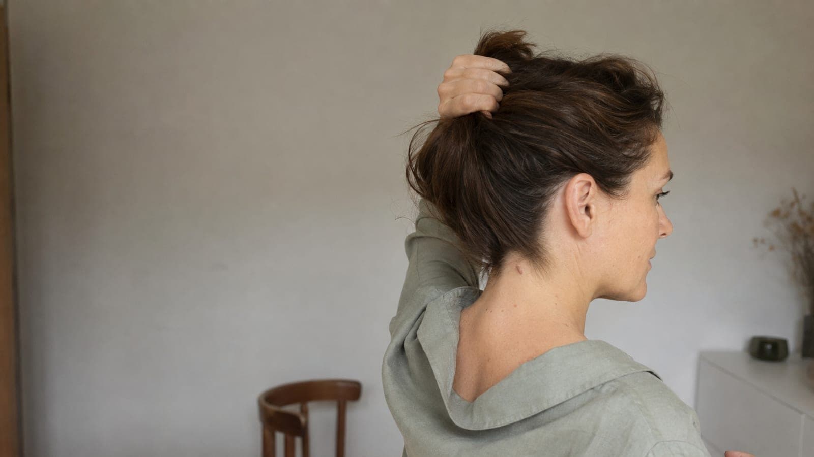
An epidermoid cyst is a benign lesion felt as a firm lump under the skin, most often behind the ear, on the neck or on the back. It grows slowly and usually does not hurt, but it can suddenly turn red, warm and painful. Squeezing must be avoided, even though the internet is full of such videos. Below we describe what a cyst actually is, why it must not be squeezed, how removal proceeds, what it is sometimes confused with and how much the procedure costs.

What a sebaceous cyst is
A sebaceous cyst is a retention cyst of a sebaceous gland. It forms when the opening of the gland becomes blocked — the sebum has no outflow, so it collects under the skin inside a sheath called a capsule. The result: a spherical, elastic lump, usually from a few millimetres to a few centimetres. Sometimes a dark point can be seen at its top — the plugged opening.
Its favourite places are the areas rich in sebaceous glands: the hairy scalp, the face, the area behind the ear, the neck, the back, and in men also the scrotum.
In itself a sebaceous cyst is a benign lesion. The problems start from two directions: it grows (and a large sebaceous cyst in a visible place gets in the way), or it becomes inflamed.

Why you must not squeeze it
Squeezing a sebaceous cyst removes only the contents, while the capsule stays under the skin — and goes on producing. The lesion almost always comes back, often larger. On top of that, squeezing it at home is a straight road to infection and inflammation: the cyst becomes painful, reddened, and an abscess may form that requires urgent incision.
For the same reason ointments and home compresses do not work — they may soothe the irritation, but they will not dissolve a cyst with a capsule.
How a sebaceous cyst is really removed
Effectively — that is, so that it does not come back — a sebaceous cyst is removed surgically, in its entirety, together with the capsule. The procedure takes place under local anaesthesia and lasts from a dozen to several dozen minutes; the wound is closed with precise sutures, and the excised lesion can be sent for histopathological examination.
With small lesions the CO₂ laser is also an option — the doctor decides on the method after examining the lesion. The rule stays the same whatever the method: if the capsule is not removed in its entirety, or if it ruptures during the procedure, the lesion may come back.
One important rule of sequence: an inflamed sebaceous cyst is first calmed down (sometimes an incision and an antibiotic are necessary), and the planned removal together with the capsule is performed after the inflammation has subsided. That is why with a painful, red lump it is not worth waiting for it to "go away on its own" — the sooner the doctor sees it, the simpler it is.
A few words about how this looks from the patient's side. The local anaesthetic is given by injection around the lesion — it stings for a dozen or so seconds and that is the most unpleasant moment of the whole procedure. The incision is usually made along the natural lines of the skin, so that the scar is as inconspicuous as possible, and the stitches are removed after several days to a couple of weeks, depending on the area. After a properly performed procedure the scar is linear and fades with time — but it always forms, which is worth knowing when deciding to remove a lesion that is not causing any trouble.
A sebaceous cyst or something else?
Things that look similar to a sebaceous cyst are a lipoma, a fibroma or an enlarged lymph node — and the distinction matters, because each of these lesions requires different management. The assessment takes the doctor a few minutes. Lesions on the eyelids call for particular caution — do not experiment there at all.
There are also situations in which a lump under the skin needs not so much removing as examining. Book an appointment sooner if the lesion is growing markedly faster than before, is hard and cannot be moved over the underlying tissue, has ulcerated or is bleeding, or if many cysts appear at the same time in a young person. In such cases the standard is to excise the lesion whole and send it for histopathological examination.
How much it costs
Surgical excision of a lesion costs from 1000 zł (lesions on the face from 2000 zł, on the eyelid from 2500 zł). You will find the details on the page about surgical removal of skin lesions — the procedure is performed by a surgeon in Krakow and Warsaw.
W naszych klinikach
What we can do about it
Zabiegi
Nie wiesz, który zabieg będzie właściwy? Surgical consultation — dobierzemy metodę do Twojej skóry.
Sources
Last updated: 14 sierpnia 2026