
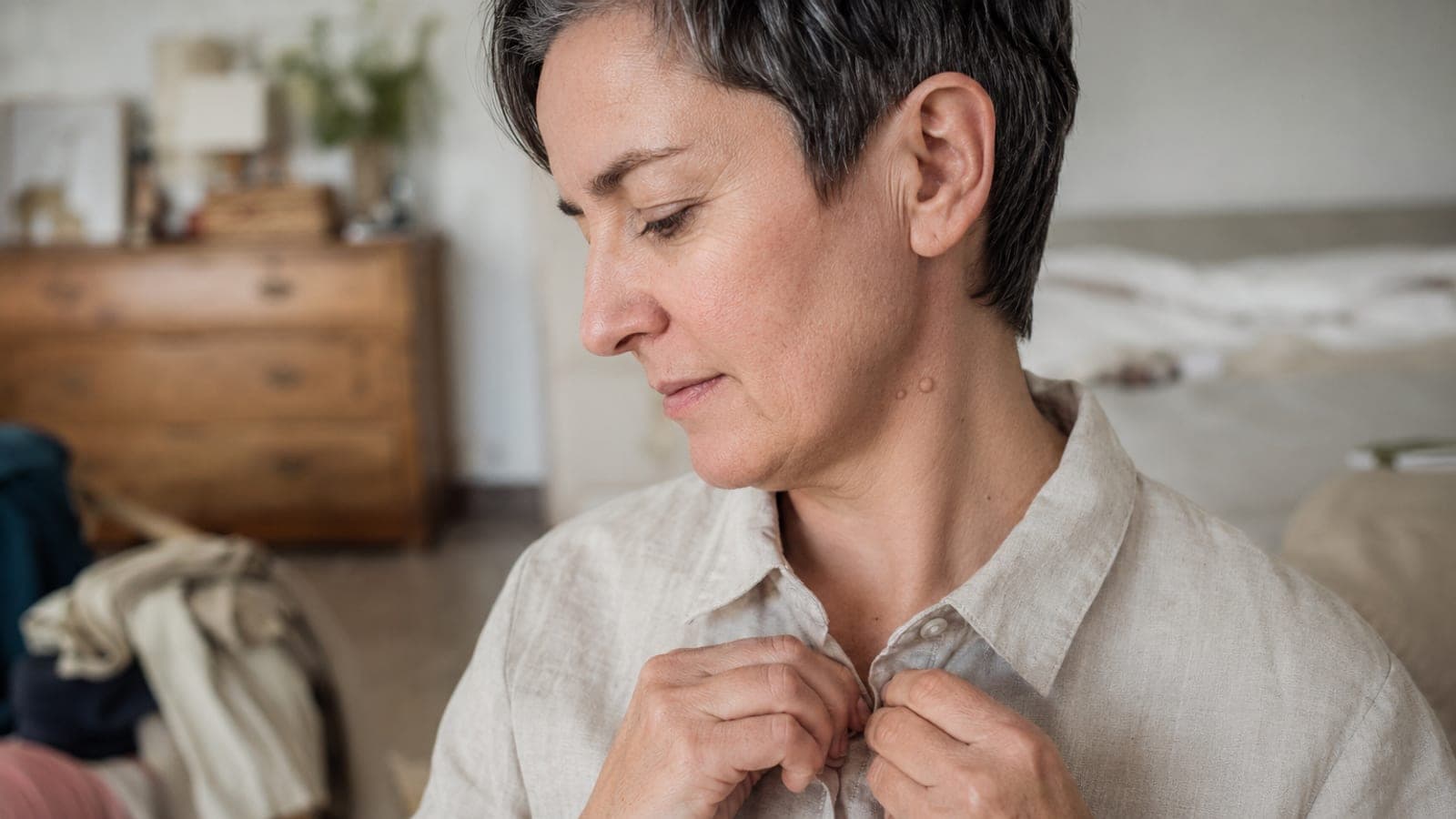
A fibroma is a benign skin lesion. It does not metastasise, and it is usually removed for cosmetic rather than medical reasons. It comes in two forms that differ in appearance, cause and method of removal: the soft one, hanging on a narrow stalk, most often on the neck, the eyelids and in skin folds, and the hard one, felt as a lump under the skin. You will find here a description of both forms, the removal methods used in the clinic, a warning against home remedies and the situations in which a lesion should not be left.
A fibroma is a tumor — and that is exactly what is not worth fearing
The word "tumor" sounds alarming, so let us explain right away what this is about. A fibroma is a benign tumor, which arises from connective tissue. The cells of that tissue — fibroblasts — multiply excessively in one patch of skin, and that is how the lesion forms.
But that is where the growth ends. Fibromas do not metastasize. They are usually removed for cosmetic rather than health reasons.
There are two basic types: soft fibromas (also called cutaneous) and hard fibromas (subcutaneous). They differ in almost everything — in how they form, how they look and how they are removed. That is why it is worth telling them apart before we go any further.

Soft fibroma: a pear on a branch
The medical names are acrochordon or fibroma molle. In practice it is a lump usually 2–5 millimeters across, suspended on a narrow stalk — exactly the way a pear hangs from a branch.
The color varies: from flesh-colored to brown. The surface is usually wrinkled, the structure flaccid, hanging like a small sac. It is built of overgrown dermis covered with epithelium. Characteristically, under pressure it can often be pushed freely back into the skin.
It rarely occurs singly. Most people have anywhere from a dozen to several dozen of them.
Darker fibromas contain more melanin, the pigment that accumulates in the skin. You will find them mainly in flexural areas — under the arm, on the elbow, in the groin — and also wherever something irritates the skin every day: a collar, a bra strap, a chain. Hence so many fibromas on the neck.
They also appear on the eyelids. In this location do not experiment — make an appointment with a specialist, who will examine the lesion and decide what to do next. In our clinics we remove eyelid lesions with the methods that require the greatest precision — plasma or the CO₂ laser, as part of non-surgical removal of skin lesions.
The soft fibroma is not a congenital lesion — it develops over the course of life, and it is favoured by age, excess weight, insulin resistance and the everyday friction of the skin. It is estimated that at least one adult in four has one, and according to some papers considerably more. It can appear as early as in teenagers, but it is decidedly more common later. Their number usually grows with age, and likewise in pregnancy — a matter of changes in hormone balance.

When fibromas suddenly multiply, check one more thing
This is the passage worth remembering.
It has been found that multiple fibromas may indicate metabolic syndrome. It consists of: obesity, dyslipidemia, arterial hypertension and insulin resistance. Such a set suggests cardiovascular disease and requires treatment.
In other words: fibromas themselves are not dangerous, but they can be a signal that something is going on in the body that is worth investigating. If there are many of them and they are multiplying fast — tell your doctor.
If soft fibromas are appearing quickly, it is worth having some simple tests: fasting glucose, HbA1c, a lipid profile, and a blood pressure measurement. In studies involving people with soft fibromas, diabetes was found in more than half of them, against about one in ten in the comparison group. This is not a reason to panic — it is a reason to have, every now and then, the tests that are recommended to every adult anyway.
How soft fibromas are removed
Let us start with the fact that settles the matter: soft fibromas persist for life and do not resolve on their own. Waiting will achieve nothing. For the lesion to disappear, intervention is needed.
There are several methods and not every one suits every lesion. The doctor decides after examining the skin — a fibroma under the arm is approached differently than a lesion on the eyelid.
What we do in our clinics:
- CO₂ laser — Vaporization of the lesion with a laser beam · By far the best method — it leaves practically no scars or discoloration. Recommended to every patient because of its very high effectiveness and the lowest rate of adverse effects.
- Cryosurgery — Freezing the lesion with a stream of liquid nitrogen; the fibroma falls off on its own · In people with dark complexions it may leave local discoloration.
- Curettage — Mechanical removal of the lesion with a special curette · For lesions protruding above the skin surface, with no need for stitches.
- Plasma — Vaporization of the lesion with a stream of energy, without touching the skin · Small lesions in places that require precision.
- Surgical excision — Removal under local anesthesia with a scalpel, scissors or a disposable punch · The punch makes the wound heal more easily. The excised fragment can be sent for histopathological examination.
What you will not find with us: electrocoagulation, that is removal of the lesion with high-frequency current under local anesthesia. This method is sometimes recommended to people with dark complexions, because it leaves less discoloration than cryosurgery — which matters for lesions in visible places.
One advantage of surgical excision deserves a sentence of its own: the removed lesion can be examined histopathologically and you can be certain what you were dealing with.
One thing patients ask about most often: a removed fibroma does not grow back in the same place, but the procedure does not protect against new ones appearing elsewhere. The tendency to form them stays — especially with excess weight, with insulin resistance and in places constantly irritated by clothing. That is why in some patients we repeat the procedure every few years, and working on body weight and blood sugar acts here as prevention.
You will find the details of both routes on the treatment pages: non-surgical removal of skin lesions (cryosurgery, curettage, plasma, CO₂ laser — from 500 zł) and surgical removal of skin lesions with histopathological examination (from 1000 zł, lesions on the face from 2000 zł).
A thread, a pharmacy product, a "trick from the internet" — why I advise against it
Since a fibroma hangs on a thin stalk, it is tempting simply to tie it off with a thread or treat it with a product from the drugstore. You will find thousands of such tips online. They all share the same problem — and it is not only about the scar.
By tying off the lesion at home, you are making the diagnosis yourself. And with the naked eye you cannot tell a fibroma from other skin lesions — including ones that require a doctor's assessment and histopathological examination. By removing a "fibroma" on your own, you may remove something that should have gone under a microscope — and deprive yourself of the only material for examination.
On top of that come the mundane matters: inflammation after incomplete necrosis, the risk of infection, discoloration or a scar in a visible place. Pharmacy preparations "for warts", in turn, can be too aggressive for thin skin — especially on the neck, décolletage or eyelids.
Removing a fibroma at the office takes a few minutes and costs from 500 zł. Set against the risk of do-it-yourself experiments, that is simply cheap.
Hard fibroma: a lump that dimples under your fingers
The second family of lesions looks and behaves completely differently.
The hard fibroma — in Latin fibroma durum, in the literature also dermatofibroma — is a firm, slightly raised lump palpable under the skin. It is usually 3–10 millimeters. The color ranges from light brown to reddish brown, darker in the center and fading towards the edges.
Unlike the soft one, it can cause symptoms: it may itch and hurt slightly when touched.
There is one test you can do yourself. Squeeze the lesion between two widely spaced fingers. A hard fibroma will shrink or dimple inwards instead of pushing forward. Doctors call this the Fitzpatrick sign.
It appears most often on the lower limbs, less often on the abdomen or buttocks. Far more often in women, and most lesions develop between the ages of 20 and 50. It does not occur en masse — usually there are at most single lesions, more rarely a dozen or so.
It forms slowly, over several months. Then it may persist for years.
Where did it come from in the first place
Here we return to the leg-shaving scene.
A hard fibroma very often forms at the site of an insect bite or sting. It also arises on the basis of hair follicle inflammation or a minor injury — including after shaving, if the skin has been badly irritated.
So if you have found a lump matching this description, think back: did something bite you in that spot? Very often the answer is "yes" and everything falls into place at once.
Does a hard fibroma have to be removed
Usually not. Most often it goes unnoticed, because it causes no symptoms at all.
But if it is a nuisance — it looks unsightly, hurts or gets in the way in some other manner — removal is worth considering. There are two options:
When deciding to remove a hard fibroma it is worth counting the costs. The lesion lies in the dermis, so excision always leaves a scar — on the lower leg, which is the most common location, it heals slowly and can be more visible than the lump itself. That is why with a lesion that is symptom-free and raises no suspicion, we most often propose observation rather than a procedure.
- Surgical excision with a scalpel under local anesthesia — The method of choice when the lesion is suspicious — it allows a specimen to be taken and assessed histopathologically under a microscope. Used when the lesion raises doubts or is a nuisance — because the fact that a hard fibroma is harmless does not mean it will disappear: it rarely resolves on its own.
Home remedies are ineffective. Worse still, they can lead to even larger fibromas growing back and to symptoms becoming more severe. This also applies to attempts to remove the lesion yourself at home.
When not to put off a visit
Not everything that looks like a fibroma is one. Make an appointment with a dermatologist if:
- the lesion is getting bigger
- it has started bleeding
- it has changed color or shape
- pain, itching or inflammation has appeared that was not there before
- the lesions are multiplying fast
- the lesion is on the eyelid
- you simply are not sure what it is
It is worth knowing why exactly these signals matter. A single, dark, enlarging lesion that looks "like a fibroma" is sometimes something else — including an amelanotic melanoma or a basal cell carcinoma. That is why the doctor examines the lesion with a dermatoscope and, at any doubt, chooses a method that allows the material to be sent for histopathological examination. That is the only way to have certainty rather than a supposition.
Finally: there is nothing to be afraid of
Fibromas are harmless and usually innocuous growths. They give no cause for concern and most people carry them all their lives without paying them any attention.
It is worth keeping an eye on the lesion, though — even if you are not planning to have it removed. And with any doubt at all, show it to a dermatologist. The doctor will assess whether the fibroma requires any intervention at all, and will suggest the best solution, taking into account above all what you want.
Assessing a lesion takes a doctor a few minutes. Guessing on your own can take months.
W naszych klinikach
What we can do about it
Zabiegi
- Non-surgical removal of skin lesionsNon-surgical removal of skin lesions comprises scalpel-free methods – cryosurgery, curettage, plasma, and CO₂ laser – allowing safe removal of benign warts, fibromas, or seborrheic lesions after a medical qualification.
- Surgical removal of skin lesionsSurgical removal of skin lesions is scalpel excision of the lesion with the option of histopathological examination, providing oncological safety and allowing care for an aesthetic scar appearance.
Nie wiesz, który zabieg będzie właściwy? Dermatology consultation — dobierzemy metodę do Twojej skóry.
Sources
- Skin Tag (Acrochordon) — StatPearls.
- Dermatofibroma — StatPearls.
- Acrochordons as a Cutaneous Sign of Metabolic Syndrome: A Case-Control Study — PMC.
- Clinicopathologic Analysis of Dermatofibroma: A Retrospective Study of 165 Cases — PMC.
- Cross-sectional study of the association between skin tags and vascular risk factors in a bariatric clinic-based cohort of Irish adults with morbid obesity — PMC.
Last updated: 14 sierpnia 2026